Image Segmentation
Digitization at the Beaty often just means inputing text and numerical data to create a record. The goal is to get all of our specimens not only recorded but imaged.
Today we'll talk about imaging and how AI can use images to support databasing.
Imaging at the Beaty
Imaging at the Beaty can mean lots of different things. There are many processes and considerations: - Camera type and setup - Lighting type and setup - Staging (fixtures, color correction, measurement, etc.) - Object standardization and prep - Object size and dimensions - Object movement and indexing
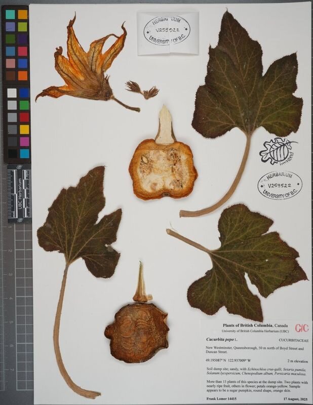
Setup for Vascular and Algae
Here is an example of the Vascular and Algae imaging setup.
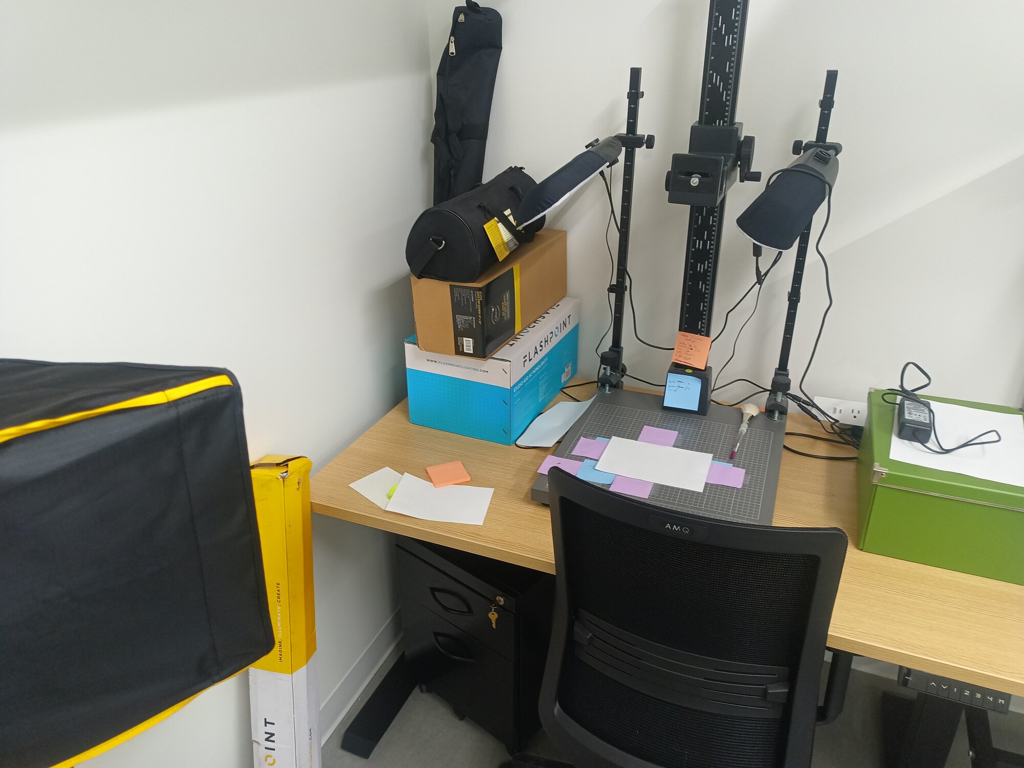
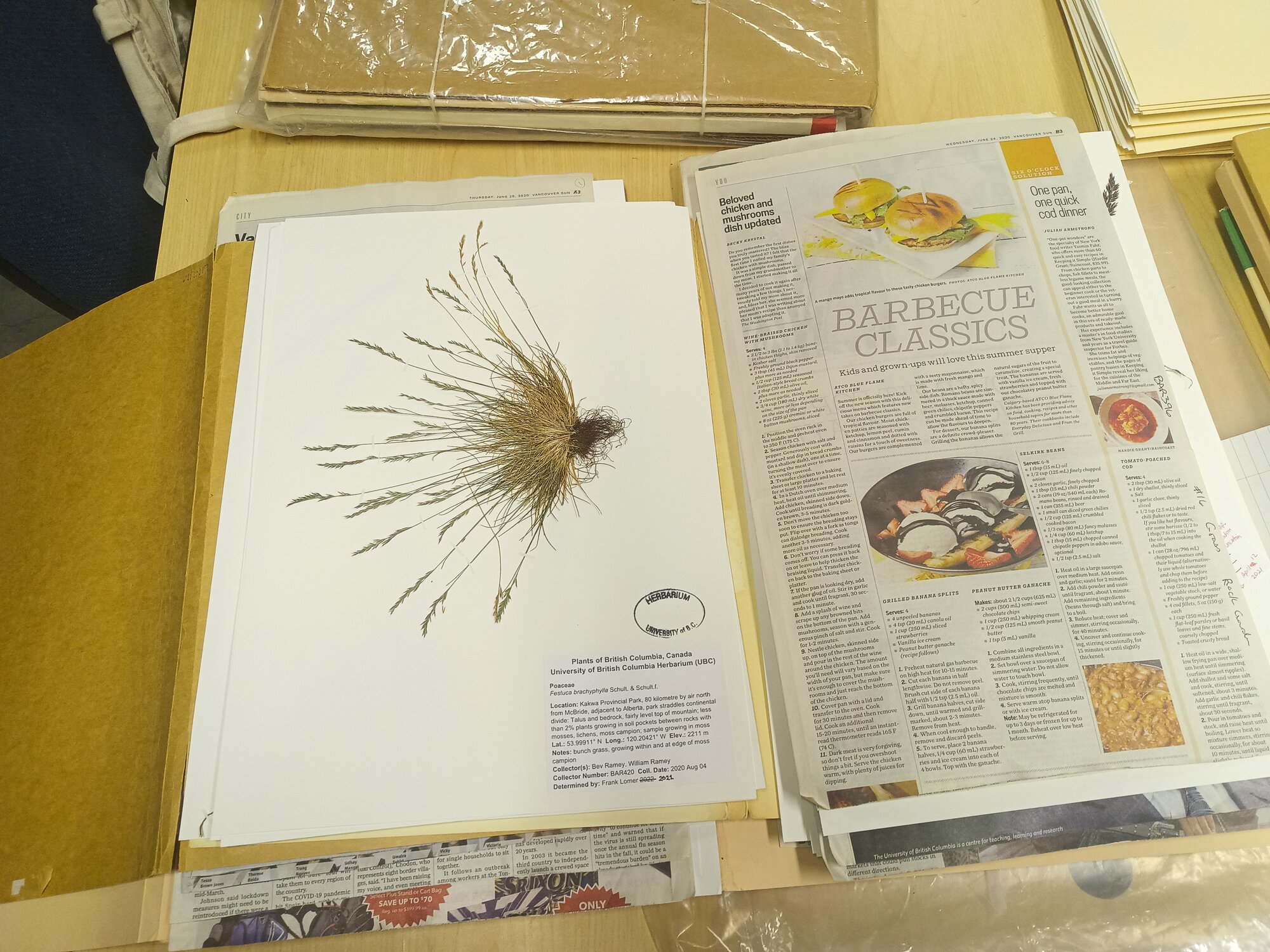
Setup for Bryophytes
Here is an example of the Bryophytes imaging setup.
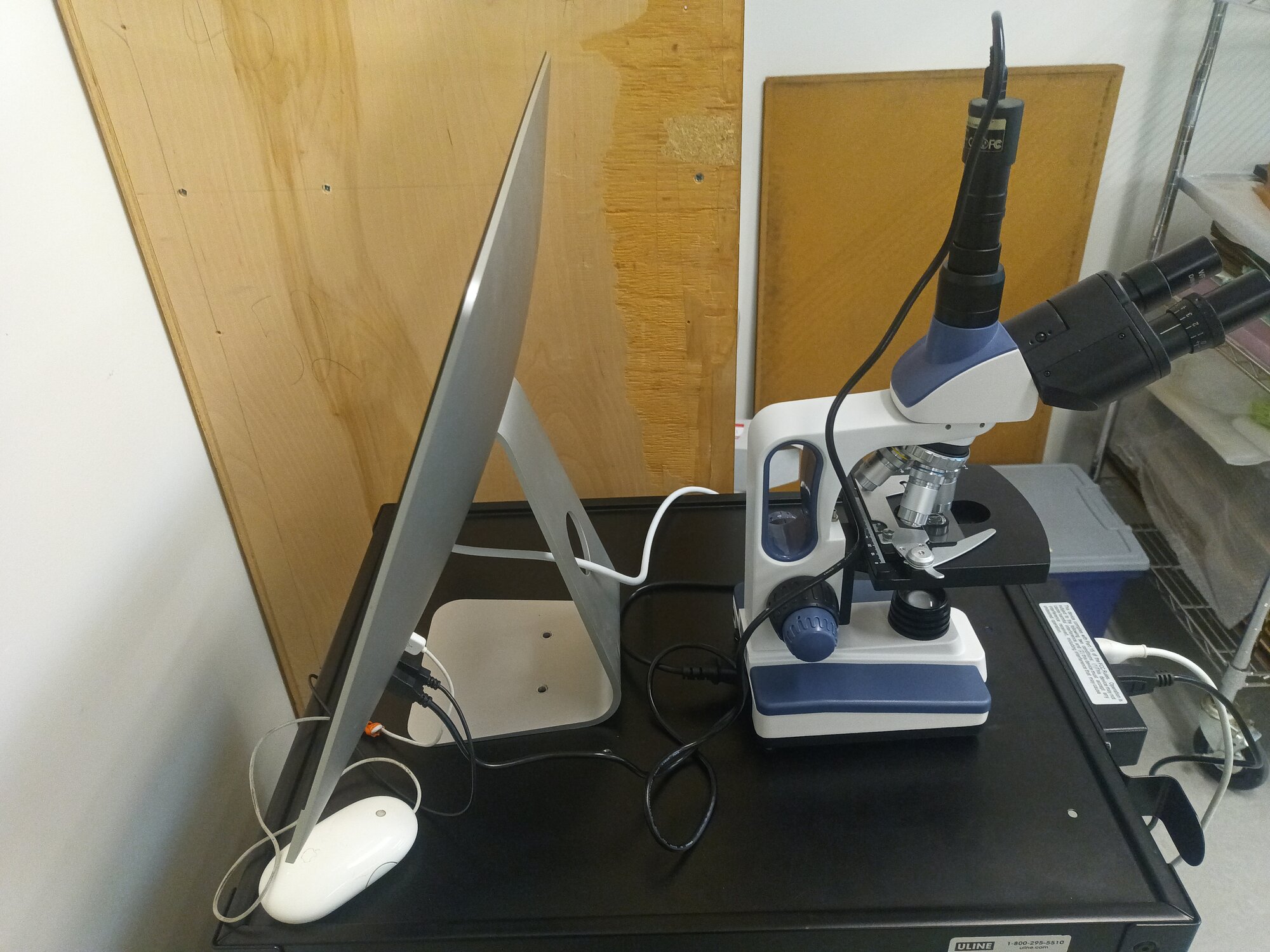
Microscopy
Entomology uses a microscope as a camera with a light ring (image similar but not exact):
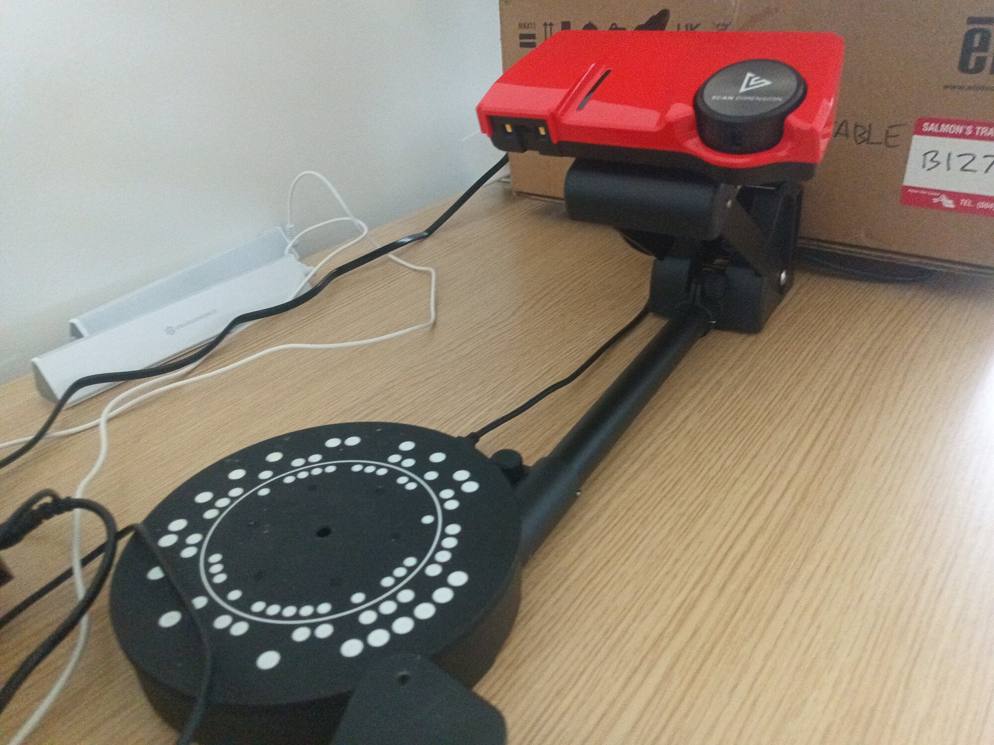
3D Imaging
Tetrapods are starting to make use of 3D imaging with combined laser and RGB setups and turntable autoindexing:
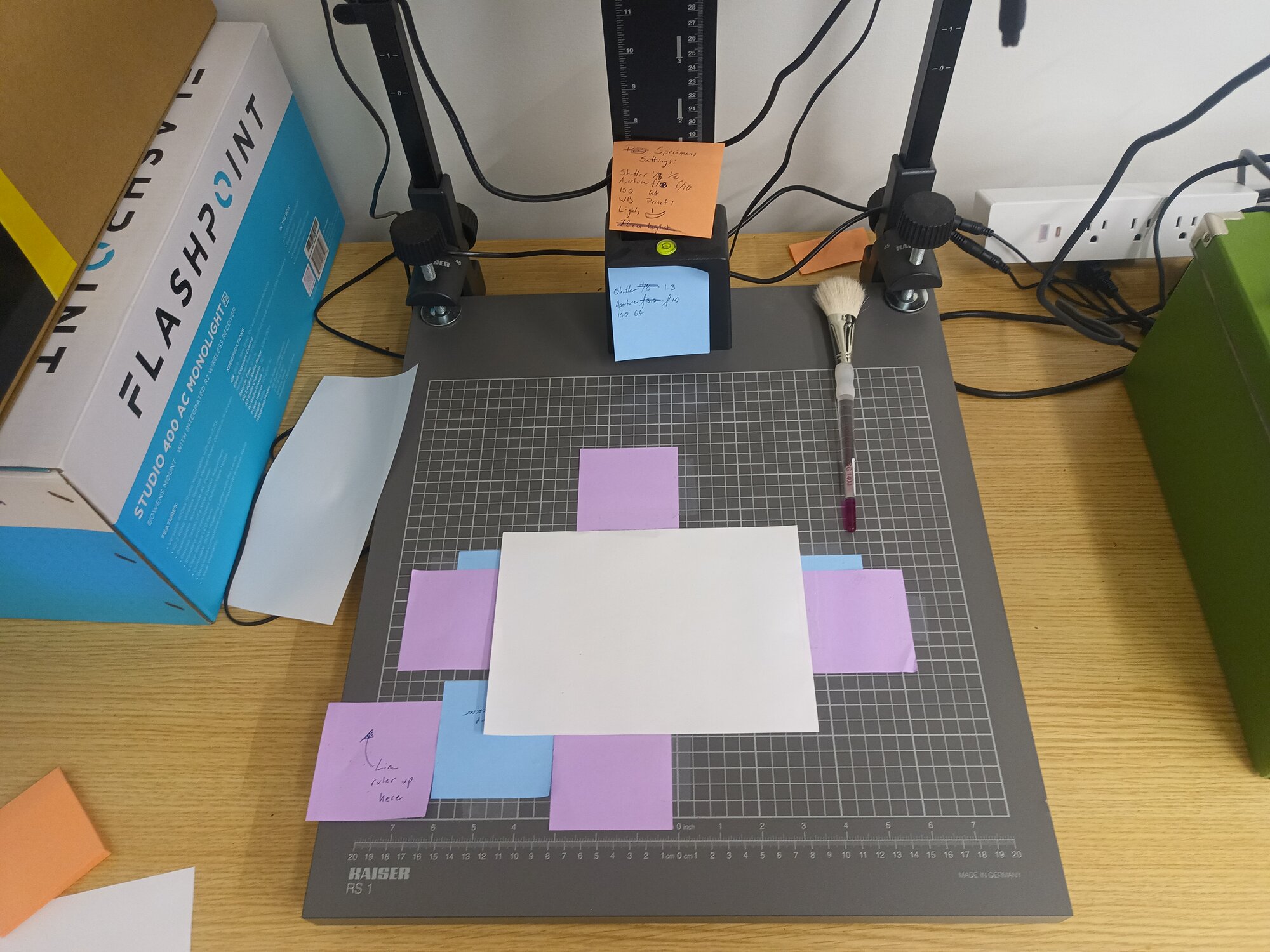
Indexing and Fixtures
Setups usually require staging
elements to index color and measurements.
Standarization and Prep
Some collections have standard sheet or package sizes:
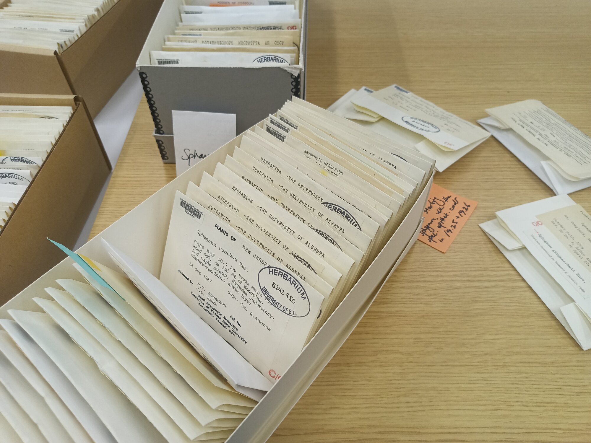 But some do not, since their specimens might be of any size.
Image Concepts: RGB
Most images are RGB raster images. That means that each pixel includes brightness values for Red, Green, and Blue LEDs that make up your screen.
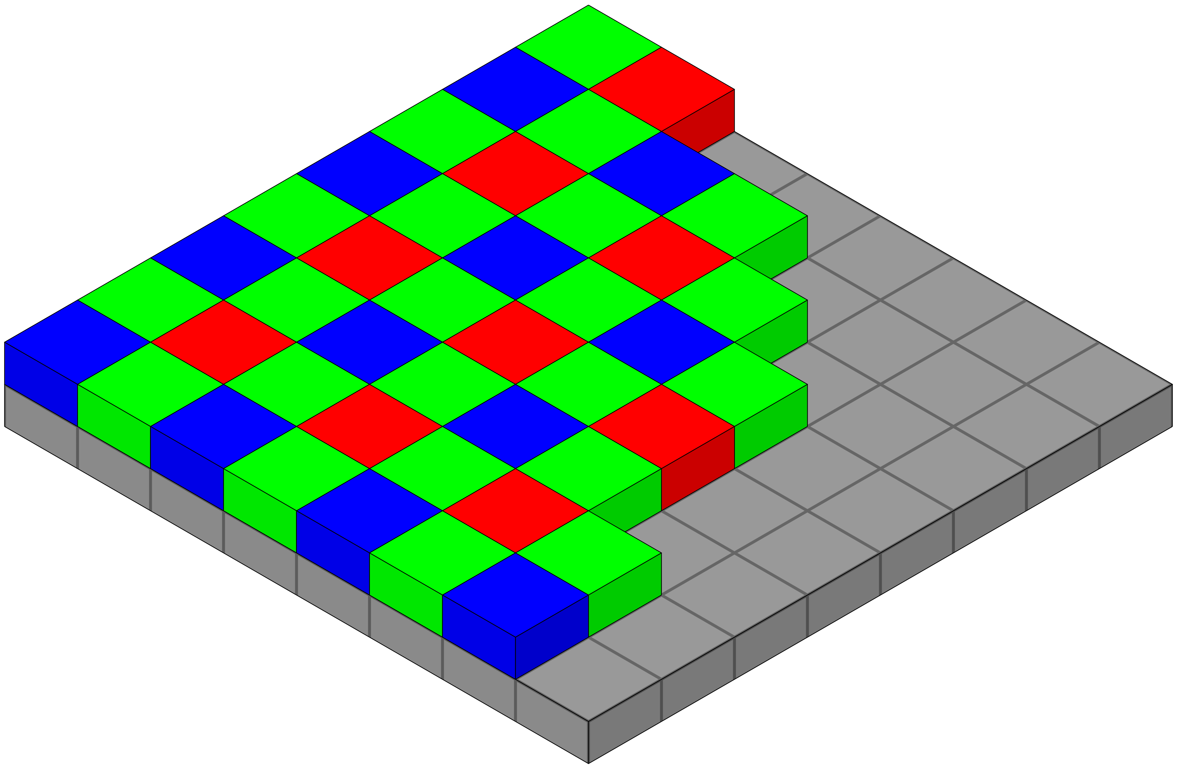
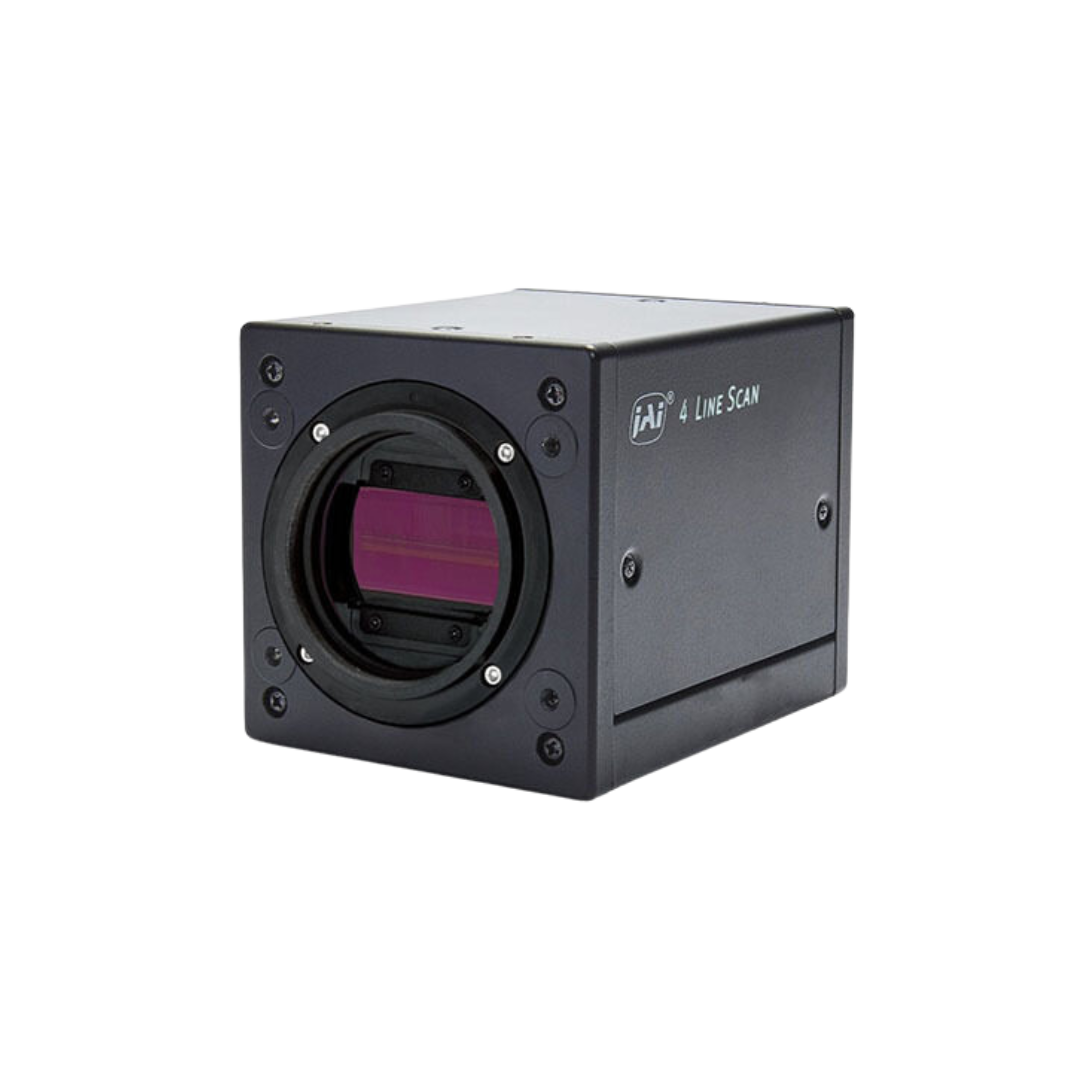
Image Concepts: Depth Map
Some images are depth maps where pixel brightness corresponds to a distance from the camera.
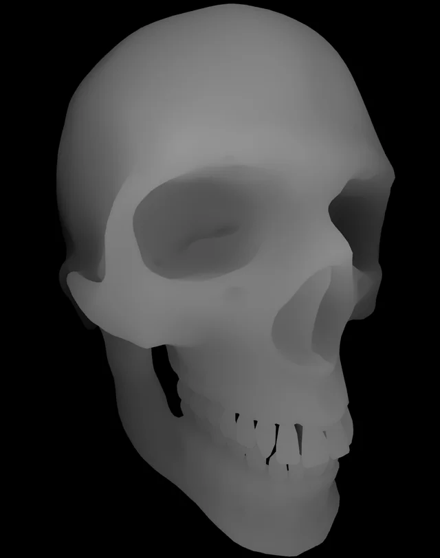
Image Concepts: Complex Imaging
Some imaging processes, like RTI, take tens or hundreds of images to be interactively reconstructed later.
https://vcg.isti.cnr.it/~palma/webrtiviewer/viewercoin.html
By the way, I want one of these domes! https://www.rti-dome.com/
Image Processing
With AI, we can now label sections of an image easily. Using a simple prompt like "plant specimen" or label we can get the following: